Clinical Pathology

HOMEActivities > Clinical Medicine > Clinical Pathology


1.Research Summary

In the field of clinical pathology, we conduct research mainly on digestive organ pathology using human tissues. We proceed with this research by adding immunohistological and molecular biological techniques to morphological analysis.

2.Organs used for research

3-1.Research subjects of the large intestine

3-2.Research subjects of the stomach

3-3.Research subjects of the esophagus

3-4.Liver, gallbladder and pancreas research subjects

4.Research Results

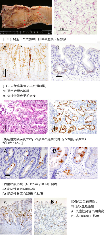
[Area] Clinical pathology (large intestine)

[Research subject]

Pathological features of inflammatory carcinogenic lesions and carcinogenic mechanism

[Description]
In long-term inflammatory bowel diseases (ulcerative colitis (UC) and Crohn’s disease), colorectal cancers occur more commonly, and this is referred to as inflammatory carcinogenesis. In our department, we conduct research on pathological features and carcinogenic mechanisms of early carcinogenic lesions mainly of UC and have clarified the following points:
① Colorectal cancers which develop in UC show histological features different from those of usual colorectal cancers: higher frequency of poor differentiation, signet-ring cell carcinomas and mucinous carcinomas; some cancers show a tendency toward dedifferentiation in the mucous membrane; many cancers show infiltrative growth in deeper sites than the submucosal layer.
② Many cancers are microscopically flat and the boundary with the surrounding mucosa is not clear, which makes it difficult to detect them endoscopically.
③ Even early carcinogenic lesions (intramucosal tumors) show characteristic histological findings, such as surface differentiation and abnormal cellular differentiation, different from those of usual colorectal cancers.
④ In early carcinogenic lesions, a reverse of the phenomenon in the proliferative zone observed in usual colorectal cancers ( the proliferative zone exists deeper in the mucous membrane in the normal colonic crypt, but it exists dominantly in the surface layer of the mucous membrane in tumors) does not occur.
⑤p53 gene abnormalities are involved, starting early in the process of carcinogenesis.
⑥ In inflammatory carcinogenic lesions and background mucosa, transformation of mucous cells from intestinal type to gastric type occurs.
⑦ In inflammatory carcinogenic lesions and background mucosa, DNA damage (double-strand break) is frequently observed.
In recent years, the frequency of inflammatory bowel diseases has also been increasing in Japan. With this, cases suffering from long-term inflammatory bowel diseases have been increasing in number, and this necessitates the development of biomarkers for the early detection and precise pathological diagnosis of inflammatory carcinogenic lesions. This research is also important for addressing these clinical needs.

[Photographs]

Photographs

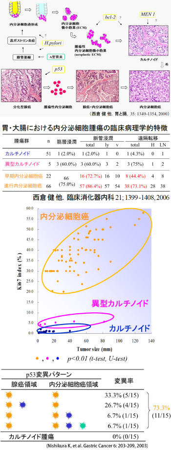
[Area] Clinical pathology (stomach)

[Research subject]

Gastrointestinal endocrine tumors

[Description]
Endocrine tumors which develop in the gastrointestinal tract are broadly divided into carcinoids and endocrine cell carcinomas. They differ not only in morphology and biological malignancy but also in histogenesis. [Nishikura K, et al., The stomach and intestines 35: 1349-1354, 2000]
Most carcinoids do not progress to muscle invasion or metastases, but endocrine cell carcinomas develop metastases at high rates from the early stage. In addition, among various tumors (carcinoids, atypical carcinoids and endocrine cell carcinomas), cellular proliferative potentials differ significantly (carcinoids < atypical carcinoids ≪ endocrine cell carcinomas). [Nishikura K, et al., Clinical Gastroenterology 21; 1399 -1408, 2006]
Histogenesis of carcinoids (cases with type A gastritis): Hypergastrinemia occurs secondary to reduced gastric acid secretion associated with fundic gland atrophy, endocrine cell hyperplasia within the affected crypts occurs (via the trophic action of gastrin), and, furthermore, the ECM is formed outside of the duct, which becomes a tumor leading to carcinoid formation. [Itsuno M, et al. Cancer 63: 881-890, 1989]
Histogenesis of endocrine cell carcinomas: A transition from carcinoids is rare, and endocrine cell carcinomas occur with preceding neoplastic endocrine cells with the adenocarcinomas as the site of origin. The bases for this are as follows:
① Endocrine cell carcinomas and carcinoid tumors seldom coexist.
② Adenocarcinomas (well- to moderately-differentiated) are associated with 78% of early endocrine cell carcinomas (14/18) and 63% of advanced endocrine cell carcinomas (27/43).
③ Most adenocarcinomas associated with endocrine cell carcinomas are intramucosal, and in 78% cases (32/41) the endocrine cell carcinoma segments continuously extend from these adenocarcinomas.
④The common p53 mutation pattern, shared between the adenocarcinoma regions and the endocrine cell carcinoma regions, is frequently observed. [Iwabuchi M, et al. Japanese journal of cancer clinics 29: 92-93, 1983] [Nishikura K, et al. Gastric Cancer 6: 203-209, 2003]

[Figures and Tables]

Figures and Tables

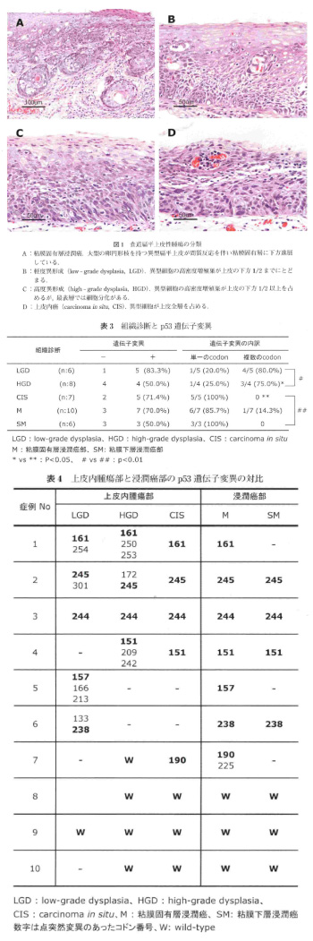
[Area] Clinical pathology (Esophagus)

[Research subject]

p53 gene mutations in the early lesions of esophageal squamous cell carcinomas

[Description]
In esophageal squamous cell carcinoma, precancerous lesions are atypical squamous cells collectively referred to as showing the features of dysplasia, which are considered to progress to invasive carcinomas through the process of carcinoma in situ (CIS). Dysplasia is broadly divided into low-grade dysplasia (LGD) and high-grade dysplasia (HGD), but it is not clear which of the two is an early carcinogenic stage for the esophageal squamous epithelium. In order to clarify the above, we focused on the p53 gene abnormalities frequently observed in esophageal squamous cell carcinomas, and investigated p53 gene mutations in squamous cell carcinomas and comorbid tumors in situ (LGD, HGD and CIS) as well as p53 protein overexpression. In 10 patients who underwent endoscopic resection for esophageal squamous cell carcinomas (invasive carcinoma in the lamina propria: M carcinoma in 4 patients, invasive carcinoma in the submucosal layer: SM carcinoma in 6 patients), we evaluated p53 immunostaining at sites of LGD, HGD, CIS, M carcinoma and SM carcinoma, and searched for p53 gene mutations. p53 gene mutations were observed in 83% of LGD, 50% of HGD, 71% of CIS, and 50%-75% of invasive carcinoma sites. In LGD and HGD, mutant codons were shared by CIS and invasive carcinoma sites coexisting in the same lesions except in one case, or both were wild type. From the above, it was estimated that LGD and HGD showed histogenetic continuity with CIS and invasive carcinoma sites in the same lesions. Gene abnormalities occurred in a single mutant codon in CIS and invasive carcinoma sites in the same lesions, but diversity was observed in mutant codons in LGD and HGD (there were multiple mutant codons). LGD and HGD were considered to consist of multiple cell populations with different p53 gene mutations, which become CIS with a uniform p53 gene mutation pattern through clone selection and then invade the lamina propria of the mucous membrane. From the results of this research, it was estimated that cell populations, in which p53 gene mutations common to both CIS and invasive carcinomas have already occurred, are already included in the LGD stage and that LGD can be positioned as an early carcinogenetic stage of the esophageal squamous epithelium. Therefore, an active clinical approach is considered to be required for LGD. [Tsuboi K. Niigata Medical Journal 124: 627-637, 2010]

[Figures and Tables]

Figures and Tables

BACK TO TOP