![]()
![]()
1.Research Summary
In the field of microscopic anatomy, we aim at visualizing and analyzing the structures and functions of cells and tissues by using various microscopic techniques. We also concurrently develop microscopic techniques for this purpose. In addition, we also attempt to obtain a comprehensive understanding of the cells and tissues by combining with techniques of molecular biology and gene engineering.
2.Three-dimensional (3D) reconstruction
We perform 3D structural analysis of tissues by reconstructing serial sections on computer software in three dimensions.
3.Scanning electron microscope
We perform 3D structural analysis of cells and tissues of each organ by using various scanning electron microscopic techniques.
4.Scanning probe microscope
An atomic force microscope is a new type of microscope which was invented in 1986. This microscope measures fine unevenness on the surface with a nanometer-scale resolution while tracing the surface of a sample with an extremely sharp needle (probe) and then creates a picture of the information.
[Area] Microscopic anatomy
[Research subject]
3D micro functional structure analysis of the Golgi apparatus by a scanning electron microscope
[Description]
From the analyses by transmission electron microscopes conducted to date,
it is known that the Golgi apparatus is made up of stacked flat cisterns
and has cis-trans polarity. However, it was difficult to analyze sterically
complex structures such as the Golgi apparatus from planar observation
employing a transmission electron microscope. Therefore, we analyze this
complex 3D micro structure of the Golgi apparatus as well as cellular functions
by using a scanning electron microscope with greater focal depth than a
transmission electron microscope which can observe samples in three dimensions.
[Photographs]
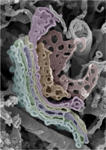
Golgi apparatus of spinal ganglion cells
[Area] Microscopic anatomy
[Research subject2]
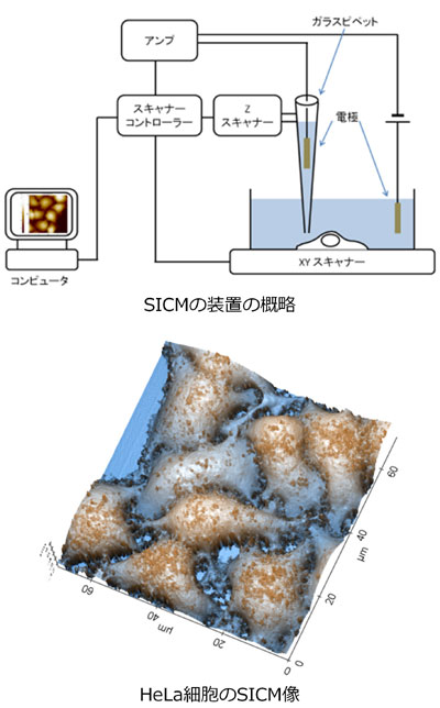
Biological applications of a scanning ion conductance microscope
[Description]
A scanning probe microscope (SPM) obtains image information by using a
sharp in-depth probe (probe) instead of a lens, by obtaining physicochemical
information generated between the probe and the sample, and by scanning
the surface of the sample while providing a form of control for it. A scanning
ion conductance microscope (SICM) is a type of SPM the biological applications
of which have been anticipated in recent years. SICM uses a micro-glass
electrode for the probe, and makes a picture of the 3D shape of the surface
without touching the sample by utilizing the change in the ion current
generated by the change in the distance between the sample soaked in liquid
and the probe. Therefore, SICM is expected to be suitable for observation
of soft biological samples in liquid, which is more similar to the physiological
environment of living things. In addition, since no specific treatment
is required for observation of samples, living samples can also be observed.
Currently, we are observing various biological samples including cultured
cells and investigating the biological applications of SICM.
[Photographs]
Please see the Microscopic Anatomy website for a detailed description of our research.