
Neurophysiology
HOME > Activities > Basic Medicine > Neurophysiology
1.Research Summary
Where does our intelligence come from? Primates such as humans and monkeys have an outstanding capacity to distinguish between visual objects, which would provide a key to the development of various cognitive abilities such as face recognition, visual imagery, and symbolic communication. We aim at clarifying the brain’s mechanisms responsible for visual cognition of objects and unraveling the mysteries underlying the intelligence of human beings by an experimental approach using primate animal models.
2.Research System
In our Lab, researchers with various backgrounds, such as animal experiments, biomedical engineering, cognitive science and clinical brain surgery, conduct interdisciplinary research by combining their knowledge and experiences. We promote personnel exchanges and actively widen the circle of collaboration through the core station of Niigata University known as the “Brain-Dream Network”, and a cross-departmental Center for Transdisciplinary Research for a project known as “Decoding and feedback of higher brain functions in primates.” We also promote collaborative studies with external research institutes, industry-university cooperation, and acquisition of intellectual properties.
3.Research Description
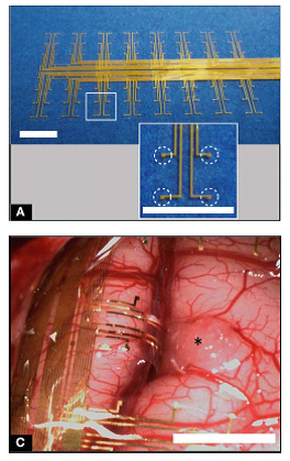
We seek the origin of human intelligence. Specifically, our research goal is to answer the fundamental question of how the distributed neural network in the cerebral association cortex underlies visual imagery and cognition, from the neuronal population level to the whole brain network level, with a millisecond time resolution. As an approach to achieving this goal, we have innovated a technique called electrocorticogram (ECoG) and specialized it for recording and decoding neural activity in the visual association cortex using primate and rodent animal models. Since 2008, we participated in the national project of “Development of Brain Machine Interface” in the Strategic Research Program for Brain Sciences sponsored by the Ministry of Education, Culture, Sports, Science and Technology of Japan. We have developed a mesh-type multichannel electrode array which widely covers the brain, in collaboration with Dr. Takafumi Suzuki at the University of Tokyo (currently at the National Institute of Information and Communications Technology) making full use of micromachine technology. This electrode has enabled to steadily record a wide range of brain electrical activities less invasively for a long time as compared with the standard penetration-type microelectrode method. Since there are holes in the gaps of the mesh, it can achieve a high adhesion with the brain, and also can be combined with penetration-type probes such as a metal microelectrode and optical fiber. Unlike the conventional scalp electroencephalogram (EEG), the electrical signals are directly measured on the brain surface with the ECoG electrode, which features a significantly elevated signal sensitivity. It also features a higher time resolution compared to other whole brain mapping techniques such as functional magnetic resonance imaging. The mesh electrode can cover a wide visual association area which corresponds to almost all areas of the temporal lobe. It can be placed not only on the external surface of the cerebral gyrus but also in the cerebral sulcus of macaque monkeys. By decoding-based analyses employing a machine learning algorithm in collaboration with Dr. Kamitani at the ATR Brain Information Communication Research Laboratory Group, it became clear that information on the visual category can be read out quite accurately from electrocorticographic signals in the inferior temporal lobe. By further advancing these findings, we expect that they may be useful for creating innovative medical technology which can decode image information in the brain and provide communication support. Meanwhile, we continue endeavoring to deepen our basic research on visual cognition and to promote a project oriented toward clarifying the neural mechanisms of higher-order functions, such as visual imagery, symbolic communication and social cognition.
4. Research Results
|
[Area] Neurophysiology |
|
|
[Research subject] Brain activity recording in the visual association area and decoding of information in the brain by electrocorticogram |
|
|
[Description] |
[Photographs]
|
Please see the Neurophysiology website for a detailed description of our research.