
Microscopic Anatomy
HOME > Activities > Basic Medicine > Microscopic Anatomy
1.Research Summary
In the field of microscopic anatomy, we aim at visualizing and analyzing the structures and functions of cells and tissues by using various microscopic techniques. We also concurrently develop microscopic techniques for this purpose. In addition, we also attempt to obtain a comprehensive understanding of the cells and tissues by combining with techniques of molecular biology and gene engineering.
2.Three-dimensional (3D) reconstruction
We perform 3D structural analysis of tissues by reconstructing serial sections on computer software in three dimensions.
- (1) 3D tissue analysis by reconstruction of paraffin serial sections
- (2) 3D structural analysis on the cell level by reconstruction of epon serial sections
3.Scanning electron microscope
We perform 3D structural analysis of cells and tissues of each organ by using various scanning electron microscopic techniques.
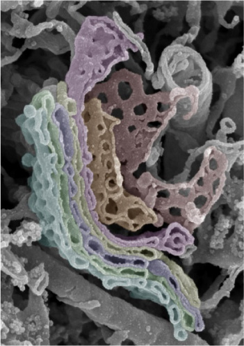
- (1) 3D micro functional structure analysis of cell organelles by the osmium maceration method
- (2) 3D structural analysis of tissues and cells by the alkaline digestion method
- (3) Analysis of vascular construction in tissues by the blood vessel molding manufacture method
- (4) Development of a real-time stereo scanning electron microscope and its biological applications
4.Scanning probe microscope
An atomic force microscope is a new type of microscope which was invented in 1986. This microscope measures fine unevenness on the surface with a nanometer-scale resolution while tracing the surface of a sample with an extremely sharp needle (probe) and then creates a picture of the information.
- (1) Analysis of biopolymers and their higher-order structures
- (2) Chromosomes as higher-order structures of DNA
- (3) Structural analysis of collagen fibrils as higher-order structures of collagen molecules
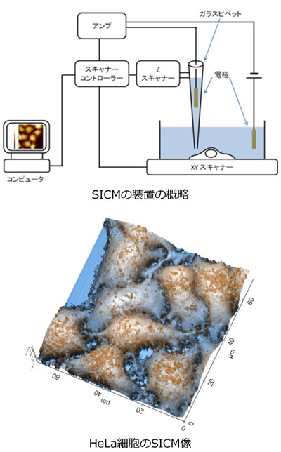
- (4) Development of a scanning ion conductance microscope and its biological applications
|
[Area] Microscopic anatomy |
|
|
[Research subject] 3D micro functional structure analysis of the Golgi apparatus by a scanning electron microscope |
|
|
[Description] |
[Photographs]
Golgi apparatus of spinal ganglion cells |
|
[Area] Microscopic anatomy |
|
|
[Research subject2] Biological applications of a scanning ion conductance microscope |
|
|
[Description] |
[Photographs]
|
Please see the Microscopic Anatomy website for a detailed description of our research.