
Radiology and Radiation Oncology
HOME > Activities > Clinical Medicine > Radiology and Radiation Oncology
1.Research Summary
Department of Radiology and Radiation Oncology consists of diagnostic radiology and radiation oncology groups. Both groups are highly active in each area of specialty as well as in a new field conglomerating their specialized abilities in each field. For the details of these research efforts, see the website of the Department of Radiology and Radiation Oncology.
2.Research Groups
- Diagnostic Radiology Group
- Radiation Oncology Group
3-1.Diagnostic Radiology Group
Research subjects
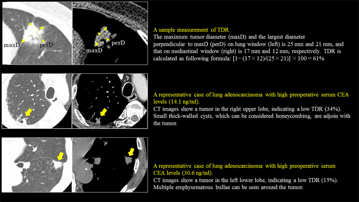
Research on diagnostic imaging for lung cancer
A wide range of research encompassing comparisons of clinical, pathological and imaging findings, time-course changes, and relationships between imaging findings and selection of therapies is performed, focusing mainly on lung cancer.
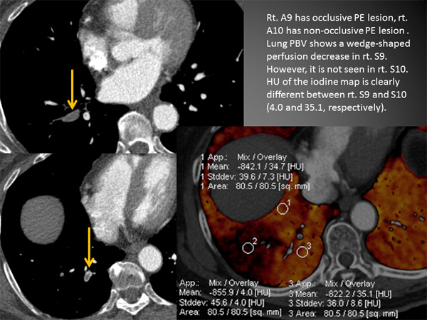
Research on the application of new diagnostic imaging procedures for circulatory
disease
Research on the application of various CT and MRI techniques
Research on therapeutic interventions using diagnostic imaging procedures
Research on RI imaging diagnosis and quantitative analysis
Research on diagnostic imaging for central nervous system diseases
addressing imaging characteristics of tumors and other diseases with recently developed technologies, such as high-spatial-resolution MRI, to promote better differential diagnosis.
3-2.Radiation Oncology Group
Research subjects
Research on the optimization of whole-brain radiotherapy in combination with stereotactic radiotherapy for metastatic brain tumors
The combination of stereotactic radiotherapy and whole-brain radiotherapy is effective for local control of brain metastases and preventing brain tumor recurrence, but conventional whole-brain radiotherapy causes the neurocognitive impairment as a late complication. Therefore, research on an optimal whole-brain radiotherapy in combination with stereotactic radiotherapy, such as a reduction in the whole-brain irradiation dose, is being conducted.
Global research on the development of new parameters of neurocognitive function on the treatment for brain tumors
Simplified tests such as the Mini Mental Score Examination have been used for evaluating cognitive function after radiotherapy for brain tumors. For establishing a more precise evaluation method, we are working on the development of a Japanese version of the cognitive function battery combining multiple cognitive function tests.
Research on the change of intratumoral blood flow during the course of stereotactic body radiotherapy for lung tumors
Intratumoral blood flow, i.e., the amount of oxygen supplied, can be estimated by perfusion CT. Perfusion CT scans before and after stereotactic body radiotherapy for lung tumors are evaluated to analyze the correlation between intratumoral oxygen concentrations and treatment response.
Research on endoscopic submucosal dissection + chemoradiotherapy for esophageal cancer
When the depth of invasion is below the mucus membrane after endoscopic submucosal dissection for superficial esophageal cancer, potential lymph node metastasis is a concern. In our department, prophylactic chemoradiotherapy is instituted in such patients to examine the efficacy of recurrence prevention.
Research on outcomes and quality of life (QOL) after intensity modulated radiotherapy and brachytherapy for prostate cancer
In our department, external irradiation (intensity modulated radiotherapy), low-dose-rate (LDR) interstitial irradiation and high-dose-rate (HDR) interstitial irradiation are performed as radiotherapy for prostate cancer. Prospective surveys and research on adverse events and the effects of change of QOL after radiotherapy are carried out.
Research on the improvement of irradiation precision with imaging guidance
In our department, we achieve high-precision irradiation by correcting
irradiation positions for each treatment session with imaging guidance
using the cone-beam computed tomography (CBCT) system during intensity
modulated radiotherapy (IMRT) for prostate cancer and stereotactic body
radiotherapy for lung tumors.
We accumulate and evaluate data on shift during treatment and conduct analyses
for improving irradiation precision.
4.Research Results
|
[Area] Radiology (medical diagnostic radiology) |
|
[Research subject] Research on diagnostic imaging for lung cancer |
|
[Description] |
|
[Photographs]
|
|
[Area] Radiology (medical diagnostic radiology) |
|
|
[Research subject] Research on the application of new diagnostic imaging procedures for circulatory disease |
|
|
[Description] |
[Photographs]
|
|
[Area] Radiology (Radiation Oncology) |
|
|
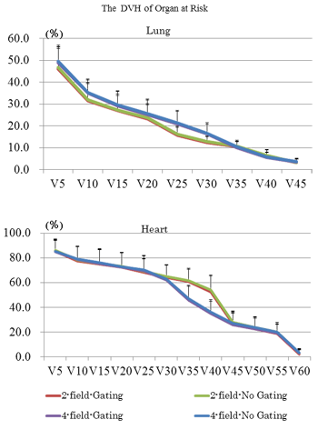
[Research subject] Research on the respiratory gating intermittent radiation and multi-field radiation for esophageal cancer |
|
|
[Description] |
[Photographs]
|
|
[Area] Medical Physics (Radiation Oncology) |
|
|
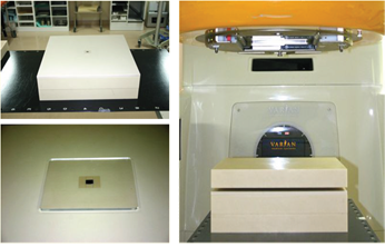
[Research subject] Evaluation of backscatter dose enhancement around dental alloy in radiation therapy for head and neck cancer |
|
|
[Description] |
[Figure 1]
The experimental setup for measuring dose enhancement. A dental alloy of 1 cm3 was embedded in a Tough Water phantom (Kyoto Kagaku) and a parallel plate chamber or a radiochromic film was placed on it. |
|
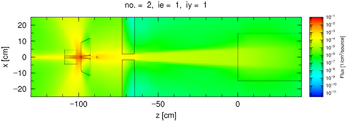
[Figure 2]
The figure describes a result of Monte Carlo calculation as an example. Target, collimator etc. in linear accelerator were modeled in Monte Carlo calculation code to calculate absorbed dose around dental alloy accurately. |
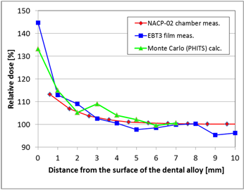
[Figure 3]
Rate of dose enhancements (vertical axis) are shown as a function of the distance from the surface of the dental alloy. The red line, the blue line and the green line stand for measured data by parallel plate chamber, radiochromic film and calculated data by Monte Carlo calculations, respectively. |
Please see the Radiology and Radiation Oncology website for a detailed description of our research.