
Genetical Skin Disease
Genetical skin disease is classified by single gene disease, multi-factor genetic disease, Chromosomal Abnormalities, and Mitochondrial genetic disease. Single gene disease occurs when some genetic mutation happens in a gene. In the dermatological field, there are two types; symptomatic, which lesions merge with skin or organs other than mucosa, and non-symptomatic, which has symptoms with skin or mucosa only.
Symptomatic Single Hereditary Skin Disease, such as Kin Fibrosis, Nodular sclerosis, Ehlers-Danlos Syndrome, Elastic Fibrous Pseudo-melanoma, and Fabry Diseases are Symptomatic Single Hereditary Skin Diseases. On the other hand, Non-symptomatic Single Hereditary Skin Disease, such as the angle of the disease Ringworm of fish scales)from dry skin, Blister Disease Epidermolysis Bullosa), and Pigment disorder which occur due to extraordinal in melanin production (Congenital Leukopathia on eyelid skin) are the Non-symptomatic Single Hereditary Skin Diseases.
Multi-factor genetic disease occurs due to being relevant to numerous factors including genes or environment. For instance, cleft palates and cleft lips. In the dermatological field, of cleft palates and lips are relevant with atopic dermatitis and Filler Green gene mutation.
Chromosomal Abnormalities: human beings usually have 22 regular chromosomes and 1 sex chromosome. However, if there is a part of extraordinariness in chromosomes or differences between numbers of chromosomes, Chromosomal Abnormalities develop. An example is Down Syndrome, the presence of part or all of a third copy of chromosome 21.
Mitochondrial Genetic Disease: Mitochondria is present in all cells in our body. It produces energy. Mitochondria also have a small amount of their own DNA. When genes have issues, the abilities of the Mitochondria decline.
In Niigata University, we provide care for Single Gene Disease, Chromosomal Abnormalities, and Mitochondrial Genetic Disease.
Epidermolysis Bullosa
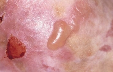
Epidermolysis Bullosa is due to having abnormalities from birth with protein that connects to the epidermis and dermis. These are single gene skin diseases, such as blisters or a sore on skin or mucosa from the slight external forces in daily life (Figure).
Usually, the proteins are stuck in between epidermis and dermis. They are formed not to dissociate from each other easily even when faced with external force. But Epidermolysis Bullosa can start to be seen when proteins aren’t produced normally. Epidermolysis Bullosa can be classified broadly into three groups by locating histologically when the epidermis and dermis dissociate; simple type, joint type, and nutritional disorder type. Then they are subdivided by the skin symptoms, genetic form or test.
The examination of the disease type uses Fluorescence Antibody Method, antibody that has recognition to find the protein between epidermis and dermis. Searching each protein condition including normal, weak, or deficit from the patient’s skin. If necessary, we will investigate genetic mutation to have accurate examination.
The Division of Dermatology in Niigata University provides the care and analysis for Epidermolysis Bullosa by cooperating with other medical facilities throughout Japan, using Fluorescence Antibody method, microscopes, and gene search. We also work together with DebRA Japan, an organization for Epidermolysis Bullosa patients and support the patients to have comfort to live their lives.